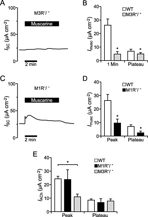
Figure 8.

Effect of muscarine on the ion transport of the tracheal epithelium of M3R−/− and M1R−/− mice. (A) Typical current observed in a M3R−/− tracheal epithelium upon application of 100 µM muscarine luminal. Application of 100 µM muscarine led to a sustained current increase. (B) The muscarine-induced current increase was significantly reduced in the tracheal epithelium of M3R−/− mice (n= 4) compared with WT mice (n= 7, P < 0.05) and the initial peak occurring in WT mice could not be detected. (C) The representative current trace shows perfusion of the tracheal epithelium of M1R−/− mice with 100 µM muscarine luminal, which resulted in an increase in the ISC that consisted of a transient peak and a lower plateau. (D) The muscarine-induced current increase occurring in M1R−/− mice (n= 4) was significantly reduced compared with WT mice (n= 7, P < 0.05 for peak and plateau). (E) The peak current increase mediated by ACh (100 µM, luminal) was significantly reduced in M3R−/− mice compared with WT controls (n= 7, P < 0.01) and not altered in the M1R−/− mice (n= 7, P= 0.76). The ACh-induced plateau current increase was not changed in M3R−/− mice (n= 7, P= 0.56) or in M1R−/− mice (n= 7, P= 0.51) compared with control ACh effects in WT mice.
