
Figure 4.
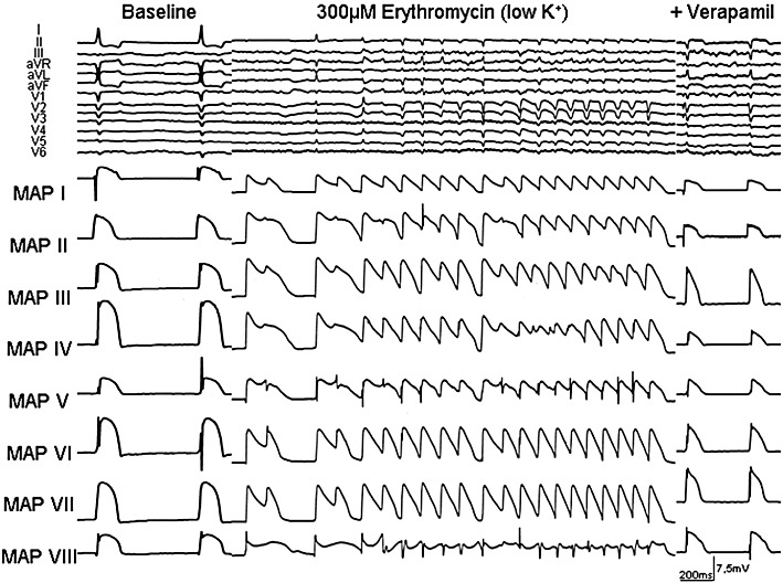
Representative example of EAD and polymorphic VT in the presence of erythromycin and after additional infusion of verapamil during bradycardia (AV block) and hypokalaemia in an isolated Langendorff-perfused heart from a CHF rabbit. ECG characteristics and MAP recordings [distribution of MAP catheters. Left heart: MAP I = base anterior, MAP IV = base posterior, MAP V = between basis and apex (posterolateral), MAP VI = between basis and apex (inferior); MAP VII = apex, MAP VIII = endocardial; right heart: MAP II = apex, MAP III = bases].