Figure 2.

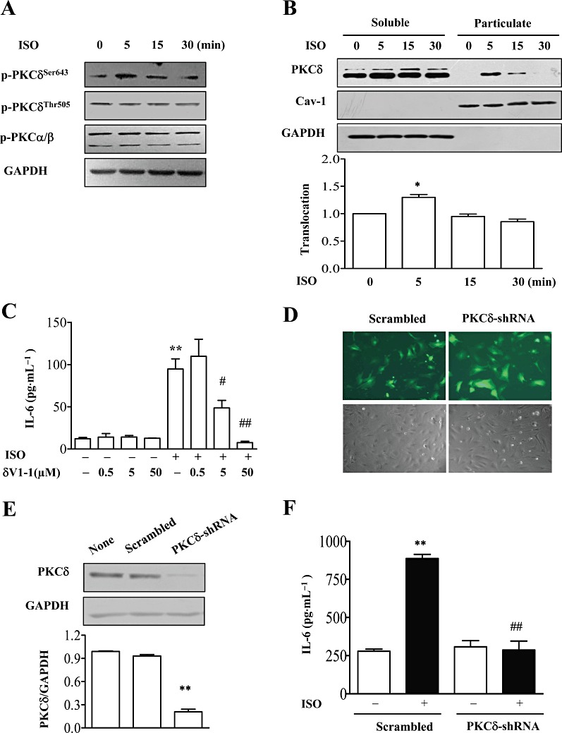
PKCδ mediated isoprenaline (ISO)-induced IL-6 production. (A) NMCFs were stimulated with isoprenaline for different times, and cell lysates were immunoblotted with antibodies against PKCδ phosphorylated at Ser643 or Thr505 (p-PKCδ), p-PKCα/βII phosphorylated at Thr638/641 and GAPDH. A representative image from three independent experiments is shown. (B) NMCFs were treated with isoprenaline (10 µM) for 5, 15 and 30 min, and cell lysates were separated into soluble or particulate fractions that were immunoblotted with anti-PKCδ antibody, anti-caveolin-1 and GAPDH. In the lower graph, mean ± SEM of data from three independent experiments. *P < 0.05 vs. value at 0 min, n= 3. (C) NMCFs were pretreated with the PKCδ translocation inhibitor (δV1-1) at various concentrations for 60 min and incubated with isoprenaline (10 µM) for 12 h. The concentration of IL-6 in cell culture supernatants was assayed by ELISA. **P < 0.01, significant effect of isoprenaline; #P < 0.05, ##P < 0.01, significant effect of δV1-1; n= 3. (D) The transfection efficiency of NMCFs infected with adenovirus expressing PKCδ-shRNAs or scrambled RNA for 48 h. Green, GFP fluorescence. (E) Effect of PKCδ-shRNAs on PKCδ protein levels. NMCFs were infected with adenovirus expressing PKCδ-shRNAs or scrambled RNA for 48 h. Cell lysates were immunoblotted with PKCδ or GAPDH antibody. Data are expressed as mean ± SEM of three independent experiments. **P < 0.01 vs. Scrambled. (F) NMCFs were infected with adenovirus expressing PKCδ-shRNAs or scrambled RNA, then stimulated with isoprenaline (10 µM) for 12 h. The concentration of IL-6 in cell culture supernatants was assayed by ELISA. **P < 0.01 vs. control, ##P < 0.01 PKCδ-shRNAs vs. scrambled. n= 3.
