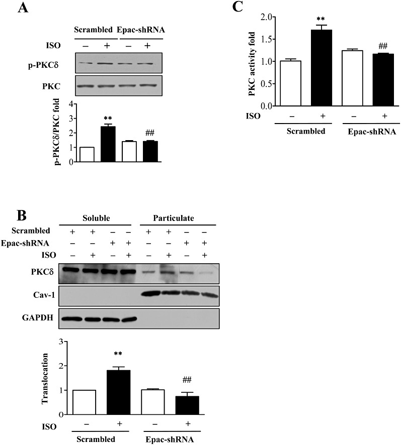
Figure 4.

Inhibition of Epac depressed β-adrenoceptor-induced PKCδ activation. (A) After knock-down of Epac1 by adenovirus, cells were stimulated with isoprenaline (10 µM; ISO) for 5 min and PKCδ Ser643 phosphorylation was determined. **P < 0.01 vs. control, ##P < 0.01 PKCδ-shRNAs vs. scrambled, n= 3. (B) After knock-down of Epac1 by adenovirus, cells were stimulated with isoprenaline (10 µM) for 5 min.. Cell lysates were separated into soluble or particulate fractions and PKCδ translocation was determined by Western blot. **P < 0.01 vs. control, ##P < 0.01 PKCδ-shRNAs vs. scrambled, n= 3. (C) After knock-down of Epac1 by adenovirus, cells were stimulated with isoprenaline (10 µM) for 5 min. and PKC activity measured by kinase assay. **P < 0.01 vs. control, ##P < 0.01 PKCδ-shRNAs vs. scrambled, n= 3.
