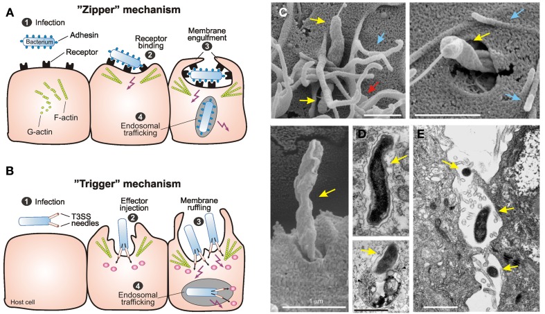
Figure 1.
Primary mechanisms of bacterial invasion into non-phagocytic host cells. Schematic representation of the two different routes of entry by intracellular bacterial pathogens. The pathogens induce their own uptake into target cells by subversion of host cell signaling pathways using the “zipper” and “trigger” invasion mechanism, respectively. (A) Bacterial gastrointestinal pathogens commonly colonize the gastric epithelium [step 1]. The “zipper” mechanism of invasion involves the high-affinity binding of bacterial surface adhesins to their cognate receptors on mammalian cells [step 2], which is required to initiate cytoskeleton-mediated zippering of the host cell plasma membrane around the bacterium [step 3]. Subsequently the bacterium is internalized into a vacuole. Some bacteria have developed strategies to survive within, or to escape from this compartment [step 4]. (B) The “trigger” mechanism is used by Shigella or Salmonella spp. which also colonize the intestinal epithelium [step 1]. These pathogens use sophisticated type-III or type-IV secretion system to inject various effector proteins into the host cell cytoplasm [step 2]. These factors manipulate a variety of signaling events including the activation of small Rho GTPases and cytoskeletal reorganization to induce membrane ruffling and subsequently bacterial uptake [step 3]. As a consequence of this signaling, the bacteria are internalized into a vacuole [step 4], followed by the induction of different signaling pathways for intracellular survival and trafficking. This figure was adapted from Tegtmeyer et al. (2011) with kind permission from Springer Publishing. (C) Scanning electron microscopy of C. jejuni 81-176 invasion. Invading bacteria (yellow arrows) were regularly associated with membrane ruffles (red arrows) and filopodia-like structures (blue arrows). This figure was adapted from Boehm et al. (2012). (D) Electron micrographs of C. jejuni–containing vacuoles (CCVs) that do not co-localize with BSA-gold (left) and CCVs that co-localize with BSA-Gold and resemble lysosomes (right, arrows) are shown. The two pictures were kindly provided by Dr. Galan (Watson and Galán, 2008). (E) The electron micrograph of translocating C. jejuni across polarized Caco-2 cells by the paracellular pathway was kindly provided by Dr. Konkel (Konkel et al., 1992b).

