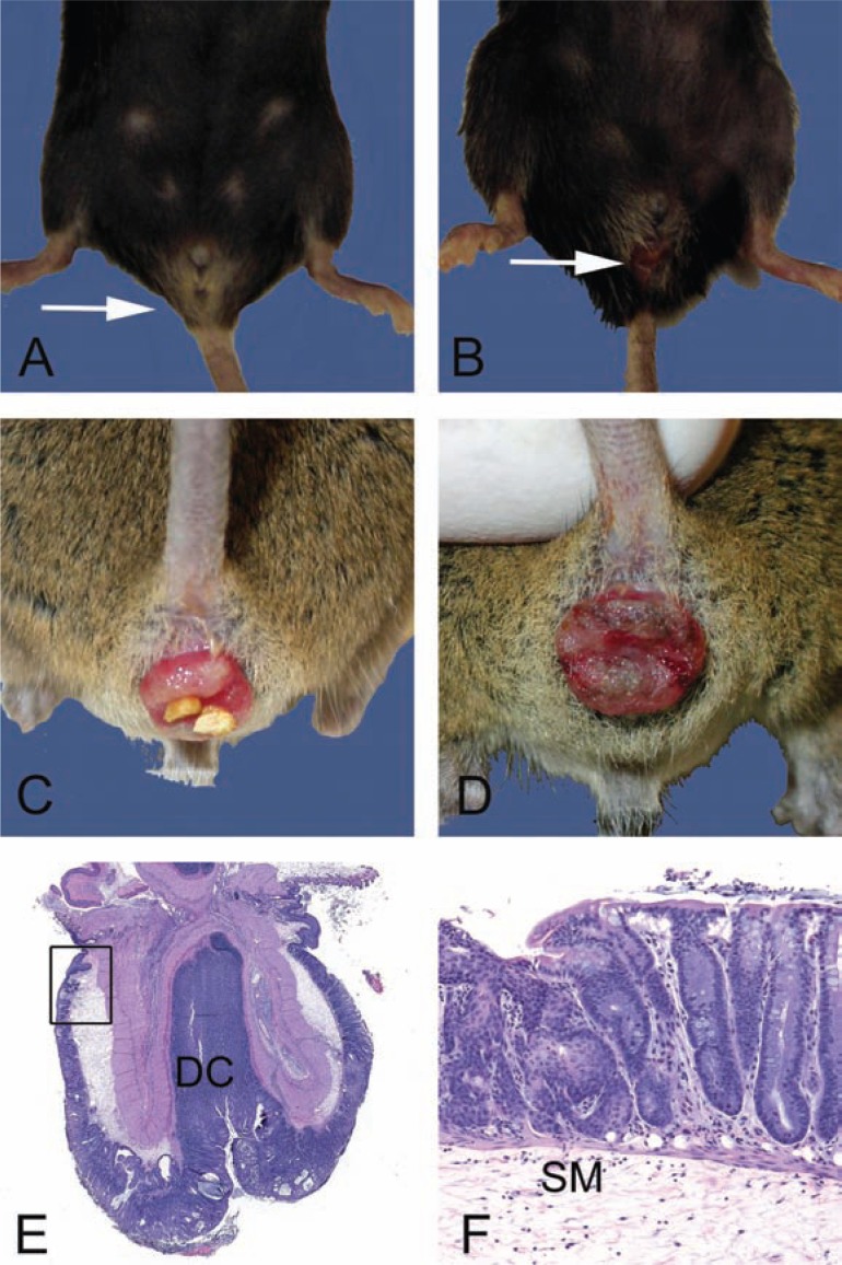
Fig. 1.
Gross and microscopic examples of rectal prolapses. A. Normal female 28-month-old B6 mouse with the anus indicated (arrow). B. Female 28-month-old B6 mouse with a mild acute rectal prolapse. Note the mild protrusion of congested and moist rectal mucosa (arrow). In female mice, rectal prolapse must be differentiated from uterine prolapse. C. Moderate to severe subacute rectal prolapse in a 4 to 6-month-old male genetically modified mouse, 129-Smad3 tm/Par/J, infected with Helicobacter species. D. Chronic severe rectal prolapse in a 4 to 6-month-old male 129-Smad3 tm/Par/J infected with Helicobacter species. The mucosa is dry and thickened with adherent crust. E. Hematoxylin and eosin-stained section of prolapsed rectum. The prolapsed mucosa is covered by a serocellular crust. Distal colon (DC) and rectoanal junction (box) are indicated. F. Higher power of boxed region in E. Squamous metaplasia (arrow) and hyperplasia of the rectal and distal colonic mucosa is common in chronically prolapsed tissues exposed to the cage environment including bedding, skin, and fecal organisms. The submucosa (SM) is markedly expanded by edema.

