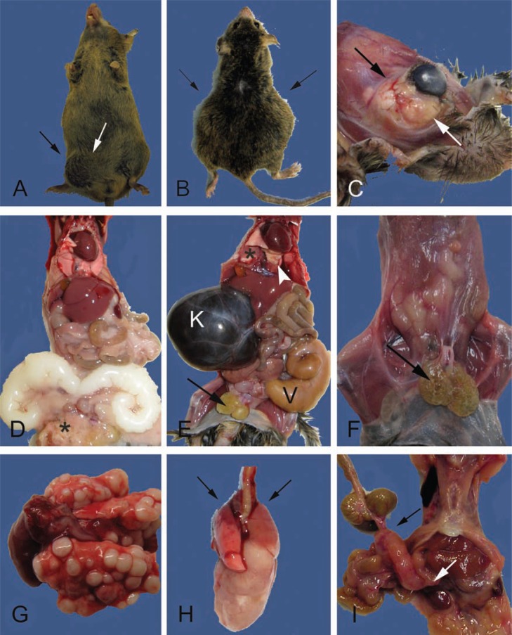
Fig. 4.
External and internal masses may be due to degenerative, inflammatory, or neoplastic lesions. A. Swelling in the lower right abdomen and the inguinal region (between arrows) are present in this 24-month-old male CB6F1. The inguinal swelling is due to an infected preputial gland. The abdominal swelling is due to enlarged, but otherwise normal, seminal vesicles (see also D). B. A 27-month-old male CB6F1 with bilateral abdominal swelling (arrows) from severe unilateral hydronephrosis and enlarged and infected seminal vesicles (see also E). C. A large multilobulated retro-orbital mass causing proptosis of the eye (between arrows). The cornea is cloudy due to keratitis (see also Fig. 3B,D). D. Internal examination of the mouse in A demonstrates bilaterally enlarged seminal vesicles, a common finding in older male mice. The seminal vesicular fluid is normally white and will discolor with infection or infarction (compare to E). Additional gross lesions are an enlarged heart with mild hepatomegaly, pneumonopathy, and the preputial abscessation (*) noted in A (between arrows). E. The internal examination of the mouse in B demonstrates severe unilateral hydronephrosis (K) and enlarged and infected seminal vesicle (V). The kidney is filled with dark brown colored fluid, a severe sequelae of obstructive uropathy (mouse urologic syndrome). Other lesions include absence of body fat, enlarged and cystic preputial glands (arrow), enlarged heart and pulmonary atelectasis (*), and light yellow firm foci (arrowhead) diagnosed histologically as AMP (see Fig. 6K,L). F. A 29-month-old male CB6F1 with bilateral cystic preputial glands (arrow), scant body fat (body condition score of 2/5), and tacky appearing tissues (mild to moderate dehydration). G. Multiple pulmonary neoplastic foci in a 30-month-old female CB6F1 mouse. This unusual presentation is more consistent with metastatic rather than primary disease. Histopathological diagnosis, however, confirmed mucinous pulmonary adenocarcinoma (see inset Fig. 5B). H. Typical presentation of a CB6F1 pulmonary adenocarcinoma from 28 to 30-month-old mice. Grossly, one large white firm mass extends from the right lung. Microscopically, there may be multiple foci of neoplasia. Cranioventral atelectasis and consolidation of the lungs is also present (arrows). I. A large mesenteric mass in the region of a cluster of lymph nodes (bracketed by arrows) from a 28-month-old B6 mouse. Differentials include lymphoma and histiocytic sarcoma. Hepatic involvement with the neoplastic process is suggested by the roughened appearance of the liver (compare to D and E) and was confirmed histologically.

