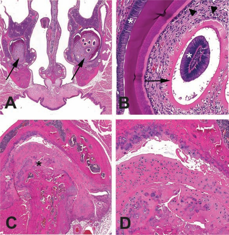
Fig. 7.
Subclinical degenerative lesions diagnosed histologically. A. Decal cross-section of the nose and incisor roots (arrows) with unilateral multiple immature intrapulpal denticles from a 28-month-old B6 mouse. B. Higher magnification of the intrapulpal denticles. Denticles with an unusual presentation of a central column of ameloblasts (*) encircled with clear space, predentin (arrow, pink band) and odontoblasts (arrowhead). Denticles may represent dysplastic tooth development. Normal position of ameloblasts (*) and odontoblasts (arrow) are also indicated. C. Osteoarthritis of the temporomandibular joint from the mouse in A with incisor dysplasia. Mandibular condyle is indicated (*). D. Higher magnification of boxed region in C. The mandibular condyle and maxillary fossal cartilages are degenerative and irregular. The joint space (*) contains free floating debris and cartilage (joint mice).

