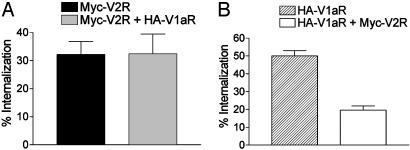
Fig. 1.
Quantitative assessment of V2R and V1aR internalization induced by AVP. HEK 293T cells expressing Myc-V2R, HA-V1aR, or Myc-V2R plus HA-V1aR were treated or not with 100 nM AVP for 30 min at 37°C to promote internalization of the receptors. The cell surface Myc epitope-tagged V2R (A)orHA epitope-tagged V1aR (B) were detected by ELISA as described in Materials and Methods. The extent of internalization was determined by measuring the cell surface receptor before and after AVP stimulation and was expressed as the loss of cell surface expression (percentage of control). Experimental conditions were established so that V2R and V1aR were expressed at ≈200 fmol per well and 100 fmol per well, respectively, whether expressed alone or in combination. All values correspond to the mean ± SEM calculated from at least five independent experiments.

