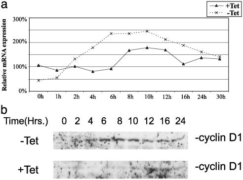
Fig. 3.
ERβ regulates cyclin D1 expression in response to E2 treatment. (a) T47D ERβ cells were spread on six-well plates at a low confluency (40%) and grown as described in Materials and Methods. Tetracycline was removed 12 h before start of treatment with 10 nM E2. Cells was harvested in TRIzol at different time points, and cDNA for real-time PCR was prepared; each point represents an average of two different cDNA preparations. (b) Whole-cell extracts were prepared from synchronized T47D ERβ cells grown on 100-mm plates as described in Materials and Methods. Proteins (50 μg) were separated on SDS/PAGE and electrotransferred to nitrocellulose membrane, and cyclin D1 protein was detected by using antibody directed against cyclin D1 (rabbit polyclonal antibody sc753, Santa Cruz Biotechnology).

