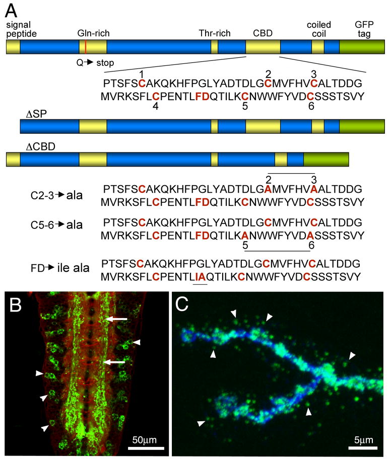
Fig 1. Transgenic constructs of the Mind-the-Gap (MTG) gene product.
(A) Diagram of MTG protein domains in wildtype and transgenic mutant variants. The Signal Peptide (SP) is deleted in the ΔSP mutant and Carbohydrate Binding Domain (CBD) deleted in the ΔCBD mutant. The red line in the Gln-rich domain indicates the Q to stop point mutation in the mtg1 null. Shown below are the three point mutant variants in the CBD domain: conserved Cys (C) residues and Phe-Asp (F–D) residues are mutated in C2-3, C5-6, and FDIA transgenic lines, respectively. (B) Representative image of elav-driven MTG::GFP expression in the wandering 3rd instar ventral nerve cord, co-labeled with anti-HRP (red) and anti-GFP (green). Arrows point to MTG::GFP within the synaptic neuropil. Arrowheads point to MTG::GFP within neuronal somata. (C) Representative image of elav-driven MTG::GFP in the wandering 3rd instar neuromuscular junction (NMJ), co-labeled with anti-HRP (blue) and anti-GFP (green). The probes were applied in the absence of detergent, to highlight labeling of extracellular proteins. Arrowheads point to MTG::GFP secreted from the presynaptic motor neuron terminal, to form extracellular puncta surrounding NMJ synaptic boutons.

