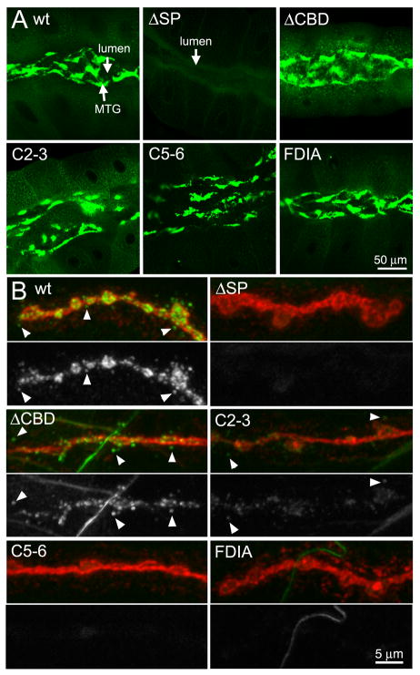
Fig 2. Expression of MTG mutant variants in the salivary gland and NMJ.
Representative images of elav-driven transgenic wildtype (wt) and mutant MTG::GFP variants in wandering 3rd instar salivary gland (A, top 6 panels), and neuromuscular junction (B, bottom panels). (A) MTG::GFP detected in the salivary gland based on native GFP fluorescence, showing protein secretion into, and accumulation within, the extracellular lumen. (B) MTG::GFP detected at the NMJ based on anti-GFP (green), with neuronal membranes co-labeled with anti-HRP (red). NMJs were labeled without detergent to highlight detection of secreted extracellular MTG::GFP. For each MTG variant, the lower panel shows GFP alone, in white. Arrowheads show secreted MTG::GFP puncta surrounding NMJ synaptic boutons.

