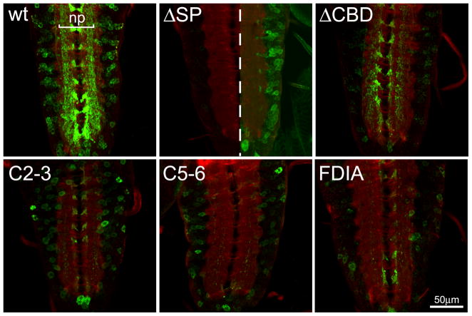
Fig 3. Expression of MTG mutant variants in the central nervous system.
Representative images of elav-driven transgenic wildtype (wt) and mutant MTG::GFP variants in the CNS of wandering 3rd instar larvae. The CNS is labeled in the presence of detergent to permeabilize the tissue, co-labeled with anti-GFP (green) and anti-HRP (red) to mark neuronal membranes. In the wt control panel, the synaptic neuropil (np) is indicated. In the ΔSP panel, the central midline dotted line divides same gain as all other genotypes (left) and increased gain to show presence of neuronal soma expression of MTG::GFP (right).

