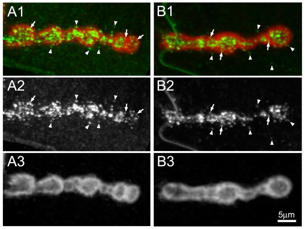
Fig 4. MTG is secreted into and beyond the subsynaptic reticulum domain.
Representative images of wandering 3rd instar NMJ synapses expressing wildtype MTG::GFP (anti-GFP, green), co-labeled with anti-Discs Large (DLG, red) to reveal the muscle subsynaptic reticulum (SSR) surrounding NMJ boutons. Detergent was used to permeabilize cells, as DLG is an intracellular, membrane-associated scaffold protein. (A1 and B1) Two example NMJ terminals showing co-labeling for MTG (green) and DLG (red) surrounding a series of adjacent synaptic boutons. (A2 and B2) Same two NMJs, showing MTG fluorescence only in white. (A3 and B3) Same two NMJs, showing DLG fluorescence only in white. Arrows indicate MTG puncta within the SSR domain defined by DLG. Arrowheads indicate MTG outside the SSR domain.

