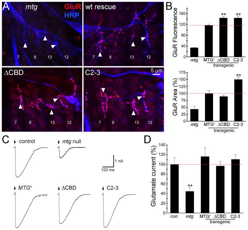
Fig 6. Mutation of CBD increases glutamate receptor abundance but not function.
(A) Embryonic NMJs (20–22 hours after fertilization) in mtg null and transgenic MTG::GFP genotypes, co-labeled for anti-HRP (blue) and anti-glutamate receptor C subunit (GluR; red). Arrowheads indicate NMJs on ventral muscles 6, 7, 12 and 13 used for quantification. (B) Quantified postsynaptic GluR fluorescence intensity (top) and area (bottom; normalized to wildtype MTG::GFP rescue) in the four indicated genotypes. The red line indicates the value of wildtype MTG::GFP rescue of the mtg1 null mutant phenotype. Asterisks indicate statistical significance (using one-way ANOVA) compared to the wildtype rescue condition (**P<0.005). Sample size is N = 5–6 hemisegments (15–18 NMJs) for each transgenic rescued genotype. (C) Representative postsynaptic currents elicited by iontophoretic glutamate application at embryonic NMJs in control (w1118; UH1-GAL4/+), mtg1 null mutant and the null mutant expressing wildtype MTG::GFP (MTG+), ΔCBD deleted MTG:GFP and C2-3 point mutant MTG::GFP transgenic conditions. Glutamate was applied as indicated (arrows) to the NMJ 6 postsynaptic domain and currents recorded in the voltage-clamped (−60 mV) muscle. (D) Quantified mean normalized glutamate response for all five genotypes. Asterisks indicate significance level compared to control (**P <0.005). Sample size is N = 7–10 embryos per each of the five genotypes.

