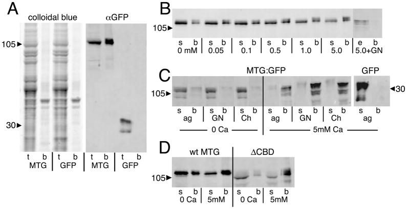
Fig 7. MTG binds to N-acetylglucosamine (GlcNAc) in a Ca2+-dependent manner.
Salivary gland lysates from wandering 3rd instar animals expressing either MTG::GFP or GFP alone assayed on GlcNAc-conjugated agarose beads. (A) Representative Western Blots of total lysate (t) and bead-bound proteins (b) separated by SDS-PAGE. Total protein (left panel) shows MTG::GFP enrichment of a small subset of proteins. Anti-GFP (right panel) shows MTG::GFP bound to beads, while GFP is found only in the lysate. (B) MTG::GFP binding is Ca2+-dependent, and competed by GlcNAc. The supernatant (s) is the lysate after incubation on beads. The bead fraction (b) is protein bound to beads. The eluate (e) is protein released from beads by 0.4M GlcNAc wash. Numbers refer to Ca2+ concentration in μM. (C) MTG::GFP binds GlcNAc (GN), GlcNAc polymer Chitin (Ch) and agarose (ag) in a Ca2+-dependent manner. GFP does not bind under any conditions (agarose shown). (D) ΔCBD also shows Ca2+-dependent binding of GlcNAc. Arrowheads indicate protein molecular weight in kDa.

