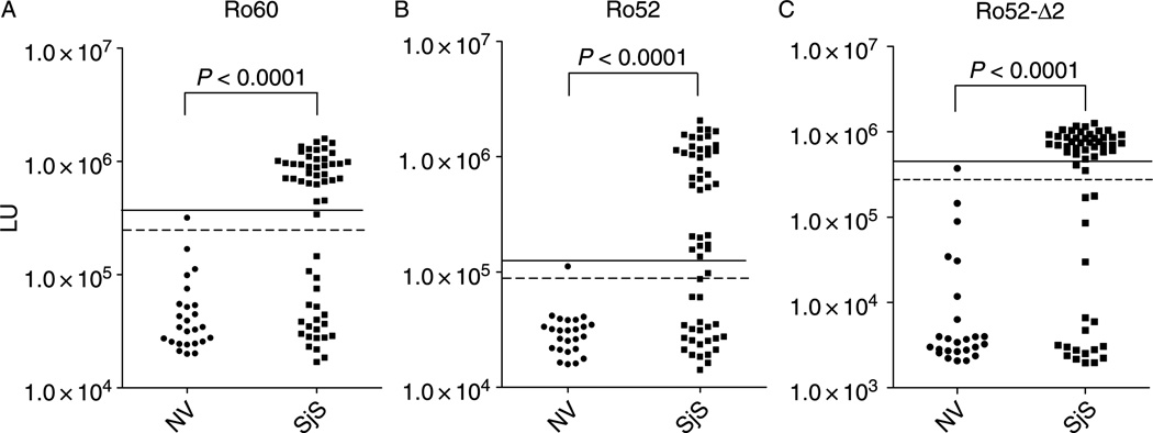
Figure 3.
LIPS detection of autoantibodies against Ro60, Ro52 and a C-terminal fragment of Ro52 in 25 normal volunteers and 57 primary SjS patients. Each circle or square symbol represents an individual normal volunteer (NV) or SjS patient sample, respectively. For determining sensitivity and specificity, the dashed line represents the cut-off level derived from mean plus 3 SD of the antibody titer of the 25 normal volunteers, while the solid line is the cut-off for the mean plus 5 SD. (A) The anti-Ro60 antibody test. (B) The anti-Ro52 antibody test. (C) The anti-Ro52-Δ2 antibody test. p-values were calculated using the Mann Whitney U-test.

