Fig. 3.
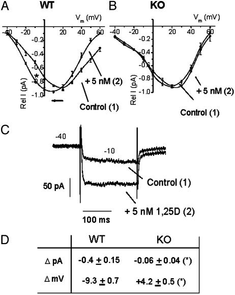
Effects of 1,25D on L-type Ca2+ channels in VDR WT and KO osteoblasts. I/Vm relations obtained for Ca2+ channel activities from VDR WT (A) and KO (B) osteoblasts, before (filled circles) and 5 min after (open circles) the addition of 5 nM 1,25D to the bath. Rel I (pA) represents the relative current value obtained for each series of 200-ms depolarizing voltage steps in relation to the maximal I (pA) value obtained for each single cell. Voltage steps were applied every second between 40 and 60 mV, from a holding potential of -40 mV. Values represent the mean ± SEM of n = 10 WT and n = 6 KO osteoblasts. Numbers in parentheses indicate the sequential order of recordings obtained from the same single cell. (C) Raw data for the typical potentiation of inward Ba2+ currents by 1,25D obtained at -10 mV from a single VDR WT osteoblast. (D) Summary of the values obtained for current amplitude increments (ΔpA) at -10 mV and shifts in I/Vm relationships (ΔmV) due to the addition of 5 nM 1,25D to VDR WT (n = 10) and KO (n = 6) osteoblasts. Data (average ± SEM) from WT and KO cells were statistically different (*, P < 0.50). Recording solutions are described in Materials and Methods.

