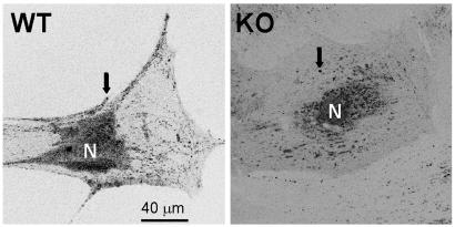
Fig. 5.
Laser scanning confocal images obtained from a VDR WT and a VDR KO osteoblast in culture for 3 weeks. Secretory granules (arrows) labeled with 1 μM quinacrine (see Materials and Methods) distribute abundantly over the cytoplasm and adjacent to the cell membrane in a VDR WT osteoblast but are scarce in the cytoplasm and virtually absent from the cell periphery in a KO osteoblast. These results are typical of ≈50 VDR WT and KO cells studied.

