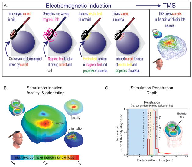
Figure 1. Foundations of stimulation.
A. Electromagnetic induction. A magnetic field (~1–4 Teslas pulsed over 0.125–1 ms, dependent on device parameters) induces an electric field that drives currents in the brain (of a magnitude of approximately 5.13 ×10⁻⁸ A/m2 in the cortex per 1 A/s steady state source, dependent on the tissue properties). Currents are carried by free charges and ions (called ohmic or resistive currents) or through the polarization of dipoles imbedded in the tissues or distributed in ionic layers surrounding the cells (called displacement or capacitive currents); see (Wagner et al. 2004). These currents are at the foundation of stimulation. B. Location, focality, orientations. The biophysical foundations can be used to determine the predicted maximum location of stimulation (*), the area of stimulation (usually ranging from ~100–200+ mm2 with a classic figure-of-eight coil with two 3.5 cm radius windings (herein defined as area from 90 to 100% of current maximum) dependent on relative head to coil placement), and the current density orientations relative to the neural tissue to be stimulated (see below). Note, circular and figure-of-eight coils are most common- whereby, figure-of-eight coils make use of the superposition of the individual fields generated by two coupled circular-coils, resulting in a more intense central-point; with circular-coils, the greatest intensity is expected to be maximum normal to the coil center-point distal from the coil face, but along the coil’s edge proximal to its face (Jalinous 1991). Of the two, the figure-of-eight coil is generally considered more focal (Ueno et al. 1988); however, the relative coil (size and position) to tissue distribution and anticipated stimulated distribution always needs to be considered, as there are conditions where circular-coils provide more focal stimulation (e.g. using small tilted circular-coils in animals (or children) with smaller heads(Wagner et al. 2007)). C. Depth of stimulation. The biophysical foundations can also be used to determine the depth of stimulation. Herein, we demonstrate the current density magnitude evaluated along an evaluation line in a healthy head, where the line is normal to the coil face and transecting all of the head tissues (note that the current density magnitude varies with the conductivity (and permittivity) of the tissues, and herein as CSF is the best conductor in the system, it demonstrates the highest current density). Generally with traditional coils, penetration is limited as the inductive magnetic fields are negligible less than a few centimeters from the scalp (for example see (Jalinous 1991)); and although one could increase the depth of stimulation with amplified source intensity, focality will conversely decrease as larger regions of tissue are exposed to increased current densities. Groups, such as Zangen’s and Davey’s, are actively pursuing improvements on these limitations (Zangen et al. 2005; Davey and Riehl 2006).

