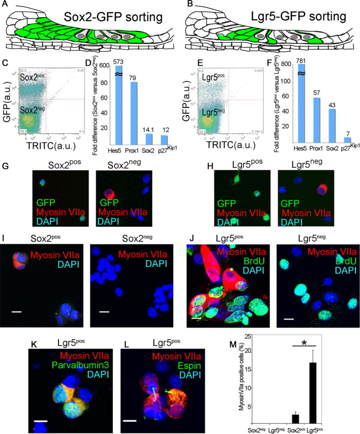
Figure 2.

Lgr5-positive supporting cells acted as hair cell progenitors in vitro. A, All supporting cells and greater epithelial ridge are labeled in the newborn Sox2-GFP mouse. B, The supporting cells and greater epithelial ridge cells shown in Figure 1 are fluorescent in the newborn Lgr5-GFP mouse. C, Sox2-expressing cells from the organ of Corti were separated by flow cytometry (Sox2pos, 23.3%). D, An enrichment in supporting cell markers in the Sox2pos fraction was seen by quantitative RT-PCR (mean, n = 2). E, Lgr5-expressing cells from the organ of Corti were separated by flow cytometry (Lgr5pos, 4.8%). F, An enrichment in supporting cell markers in the Lgr5pos fraction was seen by quantitative RT-PCR (mean, n = 2). G, The Sox2neg but not the Sox2pos fraction contained myosin VIIa-positive cells. Nuclei were stained with DAPI. H, The Lgr5pos fraction was free of myosin VIIa-positive cells which were in the Lgr5neg fraction. I, Myosin VIIa-positive cells were produced after 10 d of culture of Sox2pos but not Sox2neg cells. J, Lgr5pos but not Lgr5neg cells gave rise to myosin VIIa-positive cells after 10 d in culture, but had similar rates of BrdU incorporation. K, Differentiation of Lgr5pos cells resulted in myosin VIIa costaining with parvalbumin 3. L, Espin staining in the myosin VIIa-positive cells. M, The myosin VIIa staining in cultured Lgr5-sorted cells was 6.9 times higher than for Sox2-sorted cells. Means ± SEM are shown, n = 3.
