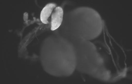
Fig. 5.
Immunofluorescence detection of Start1 protein in the PG cells. As primary antibody we used a polyclonal antibody risen against the insert amino acid sequence. The secondary antibody was labeled with Cy3. There was no noticeable difference in staining with antibodies against the complete START domain, without the transmembrane domains (data not shown). Besides the PG cells, no other tissue showed prominent staining.

