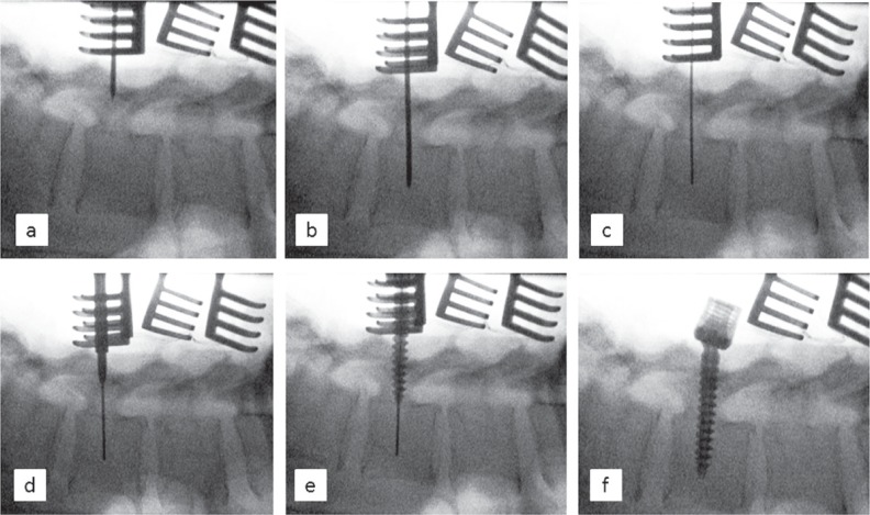
Figure 2.
Radiographic images of the process of inserting the transpedicular screw with the cannulated polyaxial screw system into spinal vertebra L4. a/b) Advancing the K-wire. c) The K-wire has been replaced by the guidance wire. d) Widening of the pedicle entrance with the cannulated awl. e) Insertion of the pedicle screw over the guidance wire. f) Final position of the pedicle screw.

