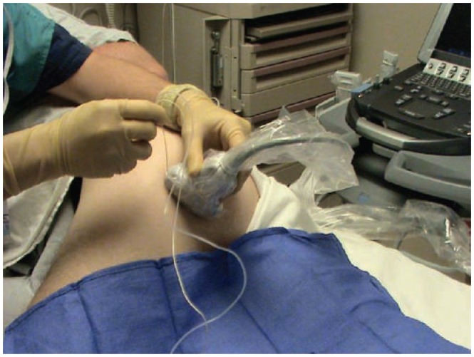
Figure 19.
The set up for a single injection infragluteal sciatic nerve block. The patient is placed in a lateral position with forward pelvic tilt. A low frequency (5–2 MHz) curved array transducer is placed on a distinct grove that can be palpated lateral to the attachment of the bicep femoris muscle on the ischial tuberosity. The image generated can be seen in Figure 20.

