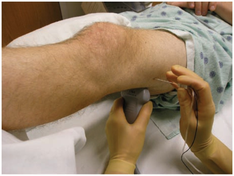
Figure 24.
The set up for an ultrasound guided popliteal sciatic block. The patient is supine with the extremity elevated. The transducer is first placed in the popliteal region and then moved in a cephalad direction until there is an optimal view of the common peroneal and tibial nerves joining to form the sciatic nerve. After measuring the depth of the sciatic nerve, the needle is advanced using an in-plane approach.

