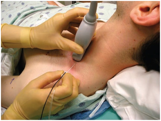
Figure 5.
The set up for performing an ultrasound guided supraclavicular brachial plexus block (BP). The ultrasound transducer is positioned adjacent to the upper border of the clavicle and the image is then optimized using the PART maneuvers (also see Figure 23). Needle advancement is from a lateral to medial direction utilizing an in-plane approach.

