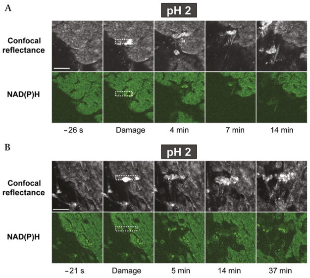
Figure 2.
Heterogeneous response to microscopic damage at luminal pH 2. The exposed gastric tissue of anaesthetised mice was superfused with a pH 2 lightly buffered saline solution during time-course imaging. Shown are results from two separate experiments in which (A) the tissue repaired in response to damage and (B) the tissue did not repair and damage continually expanded. For each experiment, representative images of confocal reflectance (upper image series) and NAD(P)H autofluorescence (lower image series) are shown. In both experiments, images were collected at the indicated times, where time zero is the time of the imposition of photodamage (in the rectangular region demarcated by the white dotted line). Images are representative of four experiments. Bar = 50 μm.

