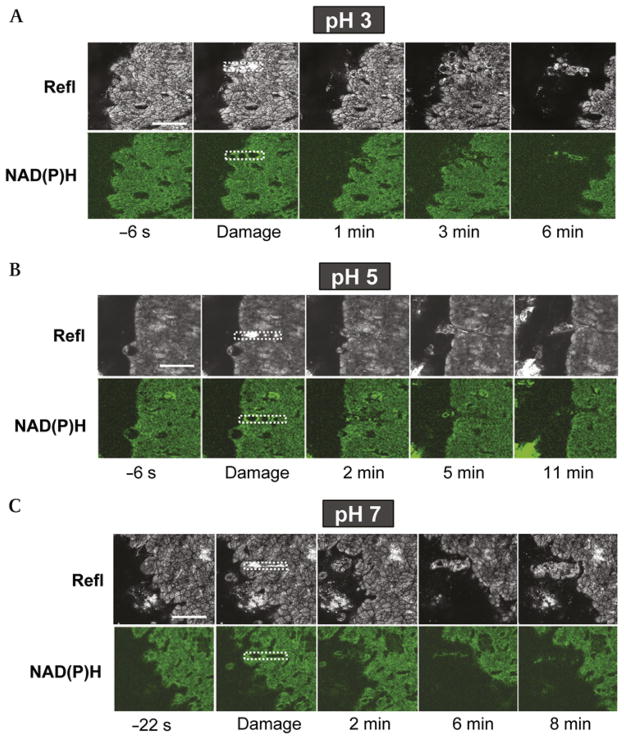
Figure 3.
Epithelial repair of microscopic damage at luminal pH 3, pH 5 and pH 7. The exposed gastric tissue of anaesthetised mice was superfused with (A) pH 3, (B) pH 5 or (C) pH 7 saline solution during time-course imaging. Otherwise, experiments were performed and results are presented as described in figure 2. Results are representative of multiple experiments performed at luminal pH 3 (n = 9), pH 5 (n = 5) and pH 7 (n = 8). Bar = 50 μm. Refl, confocal reflectance.

