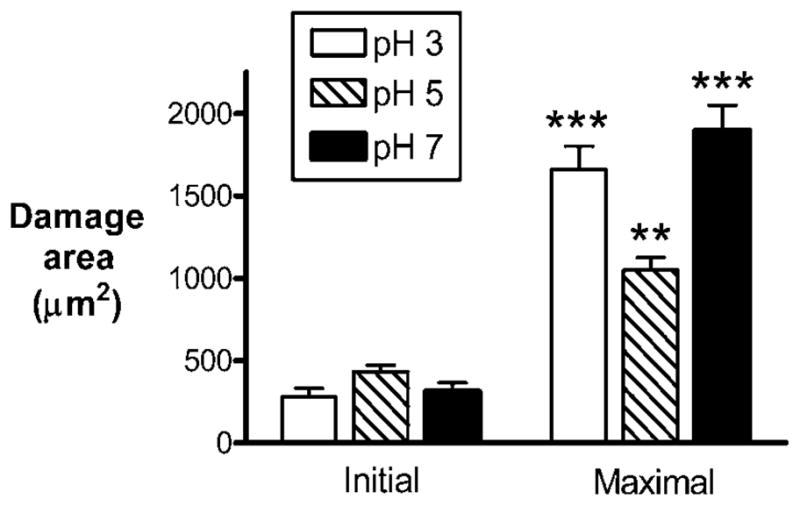
Figure 4.

Quantification of damage size at luminal pH 3, pH 5 and pH 7. The gastric mucosa of anaesthetised mice was exposed for imaging and superfused with either pH 3 (n = 9), pH 5 (n = 5) or pH 7 (n = 8) solution. Damage size was measured from images as described in Materials and Methods. The initial damage size was measured <10 s after the targeted region was exposed to repetitive scanning by high-power two-photon laser light. The maximal damage size indicates the magnitude of damage expansion (usually 3–5 min after the initial damage). Data are presented as mean±SE and analysed using ANOVA, with Bonferroni’s post hoc test. **p<0.01 and ***p<0.001 versus the respective initial damage sizes.
