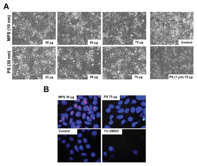
Figure 1.
Biotolerability of nanoparticles. NIH-OVCAR3 cells adherent on coverslips were incubated with 10 nm naked mesoporous silica or 30 nm COOH-polystyrene nanoparticles (at the indicated concentration) in fresh medium for 48 hours. Thereafter, the monolayers were extensively washed to remove the excess of unbound nanoparticles and (A) imaged under the phase-contrast microscope to document gross morphological alterations or cell loss, and (B) labeled with CellTracker™ to show metabolic effects.
Note: Positive control of toxicity was performed by incubating the cells with 1% dimethyl sulfoxide.

