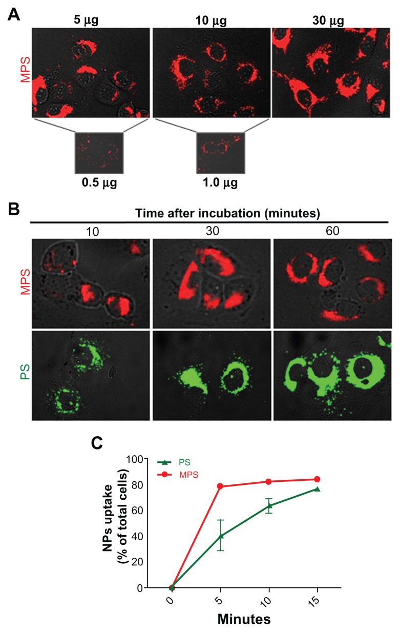
Figure 2.
Dose-dependent and time-dependent cellular accumulation of nanoparticles. (A) Cells adherent on coverslips were exposed to different concentrations of 10 nm naked mesoporous silica nano-particles for 5 minutes and imaged by fluorescence microscopy. (B) Adherent cells exposed to 10 μg/mL of 10 nm naked mesoporous silica nanoparticles or 75 μg/mL of COOH-polystyrene nanoparticles for the time indicated as imaged by fluorescence microscopy. (C) Cytofluorometric evaluation of labeled cells incubated with 10 μg/mL of 10 nm naked mesoporous silica nanoparticles or 75 μg/mL of 30 nm COOH-polystyrene nanoparticles for increasing periods of time.

