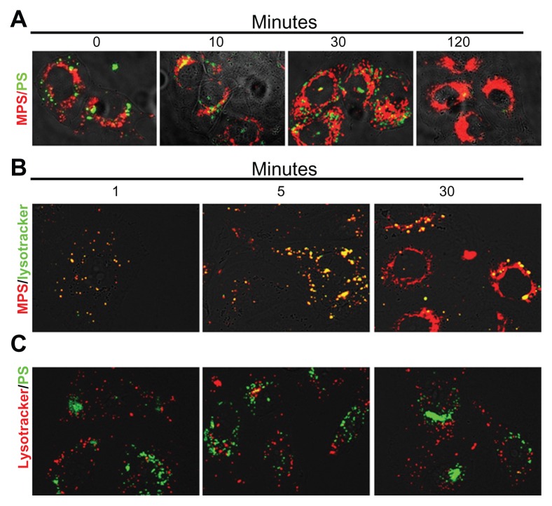
Figure 4.
Mesoporous silica and polystyrene nanoparticles take different endocytic routes and localize to distinct intracellular compartments. (A) Cells adherent on coverslips were coincubated for 5 minutes with 30 μg of 10 nm naked mesoporous silica nanoparticles and 75 μg of 30 nm COOH-polystyrene nanoparticles. The cells were then washed and imaged at 0, 10, 30, and 120 minutes of trace. (B and C) Cells adherent on coverslips were preincubated for 10 minutes with Lysotracker Green or Red, then washed and incubated with nanoparticles (as indicated), and imaged at 1, 5, and 30 minutes.

