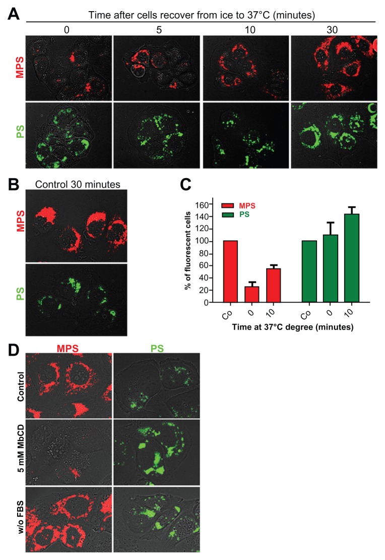
Figure 5.
Energy-temperature dependence of nanoparticle endocytosis. (A) Cells adherent on coverslips were incubated for 30 minutes in the presence of 30 μg of 10 nm naked mesoporous silica or 75 μg of 30 nm COOH-polystyrene nanoparticles at 4°C (on ice). The cells were then washed thoroughly, incubated at 37°C, and imaged at the time indicated. (B) Control cells were incubated for 30 minutes at 37°C with 30 μg of mesoporous silica or 75 μg of polystyrene nanoparticles. (C) A parallel set of cultures treated as described in panel A was used for flow cytometry evaluation of cell-associated fluorescence. (D) NIH-OVCAR cells adherent on coverslips were pulse-labeled for 15 minutes with 10 μg of 10 nm naked mesoporous silica or 75 μg of 30 nm COOH-polystyrene nanoparticles in complete or serum-free medium as indicated.
Note: Parallel cultures preincubated for one hour with 5 mM methyl-β-cyclodextrin in serum-free medium were used to assess clathrin/caveolae-mediated endocytosis.

