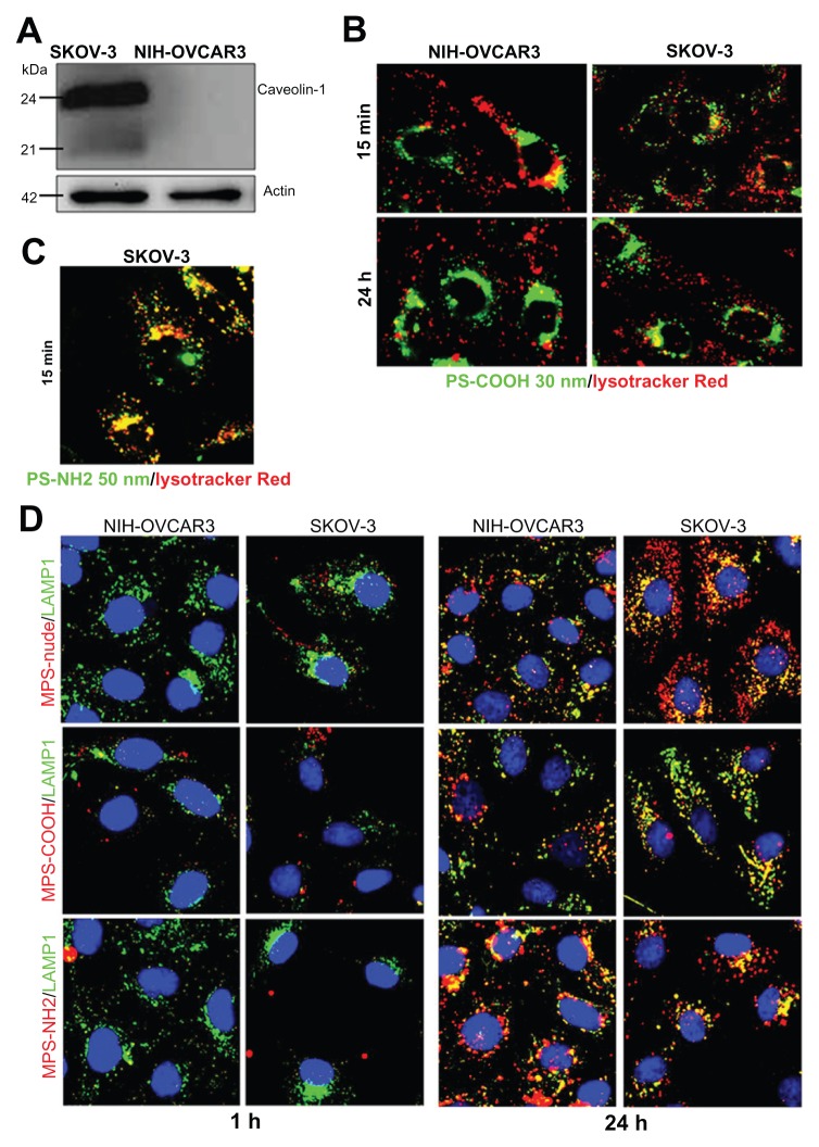
Figure 6.
Size and charged functional groups differentially affect the uptake and biocompatibility of mesoporous silica and polystyrene nanoparticles in ovarian cancer cells expressing or not expressing caveolin-1. (A) Western blotting of caveolin-1 in NIH-OVCAR3 and SKOV3 cells. The filter was reprobed for actin as a reference protein for loading of the lanes. The molecular weight of the proteins is indicated. Data were reproduced in three independent experiments. (B) Colocalization of 50 nm amine-modified polystyrene nanoparticles with Lysotracker-positive acid compartments in SKOV3 cells after an incubation time of 15 minutes with 75 μg of nanoparticles. (C) Comparison of uptake and intracellular localization of 30 nm COOH-polystyrene nanoparticles in SKOV3 and NIH-OVCAR cells after incubation times of one and 24 hours with 75 μg of nanoparticles. The 30 nm COOH-polystyrene nanoparticles showed no colocalization with the acid compartment tracer, Lysotracker Red. (D) Comparison of uptake and intracellular localization of 50 nm mesoporous silica nanoparticles functionalized or not with either COOH or NH2 groups in SKOV3 and NIH-OVCAR cells after incubation times of one and 24 hours with 20 μg of nanoparticles.
Note: Endosomal and lysosomal compartments were identified using the Lamp-1 antibody.

