Figure 5.
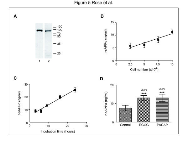
In vitrosecretion of r-sAPPα by cortical neurons in primary culture and its modulation by pharmacologic agents.A: The conditioned medium of 4 x 10⁵ neurons at six days in vitro was precipitated by a mixture of trichloraceticacid/acetone and the pellet was resupended in Laemmli buffer. An equal volume was loaded in two wells in 10% SDS-PAGE for western blotting. r-sAPPα at 100 kDa was recognized both by 22C11 (2) and by the rodent C-terminal sAPPα antibodies (1). B: Various numbers of cortical neurons (between 104 and 7 × 10⁴) were plated in 96-well plates in 0.1 ml final volume. After six days in culture, 60% of the conditioned medium was discarded and replaced by fresh medium. 24 hours later, medium was collected and 5 μl of each sample was tested in duplicate in the r-sAPPα assay. C: 5 × 10⁴ neurons were plated in 96-well plates in 0.1 ml final volume. After six days in vitro, 60% of the conditioned medium was discarded and replaced by fresh medium. Medium was collected after incubations of 3 hours, 6 hours, 9 hours or 24 hours and 5 μl of each sample was tested in duplicate in the r-sAPPα assay. D: 5 × 10⁴ neurons were plated in 96-well plates in 0.1 ml final volume. After six days in vitro, 60% of the conditioned medium was discarded and replaced by fresh medium. EGCG (30 μM) and PACAP-27 (1 μM) were added and the medium was collected 24 hours later. 5 μl of each sample was tested in duplicate in the r-sAPPα assay. ***: p < 0.001.

