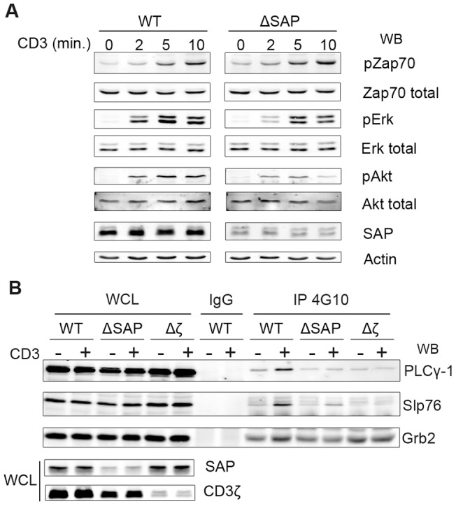
Figure 5. SAP acts in the CD3-mediated activation of Erk, Akt and PLCγ1 pathways.

A, The parental H9 cell (WT) and H9 cells, stably expressing a SAP specific Sh-RNA (ΔSAP), were stimulated with UCHT-1 for the indicated times. 25 µg of proteins were separated by SDS-PAGE and Zap70, Erk and Akt activations were revealed by Western blotting using specific phospho-antibodies. The total amount of each protein in the lysate was shown for comparison. SAP extinction was controlled by an anti-SAP immunoblotting and equal quantities of proteins in each lane were assessed by an anti-Actin immunoblotting. B, parental Jurkat cells (WT) and Jurkat clones expressing either SAP-targeting or CD3ζ-targeting Sh-RNAs (ΔSAP and Δζ respectively) were stimulated or not with UCHT-1 for 10 minutes and lysed. Proteins were immunoprecipited using a phospho-tyrosine antibody (4G10), separated on SDS-PAGE and transferred. PLC-γ1, Slp76 and Grb2 were immunoblotted using specific antibodies. This figure is representative of at least eight experiments in different clones and cell lines.
