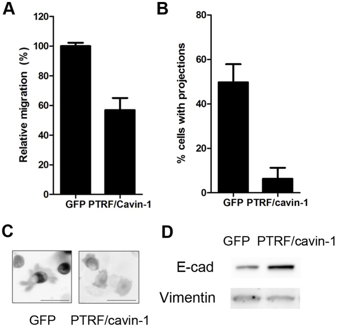
Figure 2. PTRF/cavin-1 expression in PC3 cells reduces 2D migration concomitant with reduced protrusions and mesenchymal epithelial transition.
(A) Relative migration in a wound-healing assay was assessed by time-lapse video microscopy as described in materials and methods. P<0.01 (B) Representative still images showing cell morphology. (C) Number of projections was quantitated by three individual researchers in random single cells from three independent videos (N>30 cells assessed per researcher, shown as mean ± SEM, p<0.05). (D) Total cell lysates (20 µg) from GFP or PTRF/cavin-1-GFP PC3 cells were separated by SDS-PAGE and immunoblotted using anti-E-cadherin or anti-vimentin antibodies as indicated. Data representative of 3 independent experiments.

