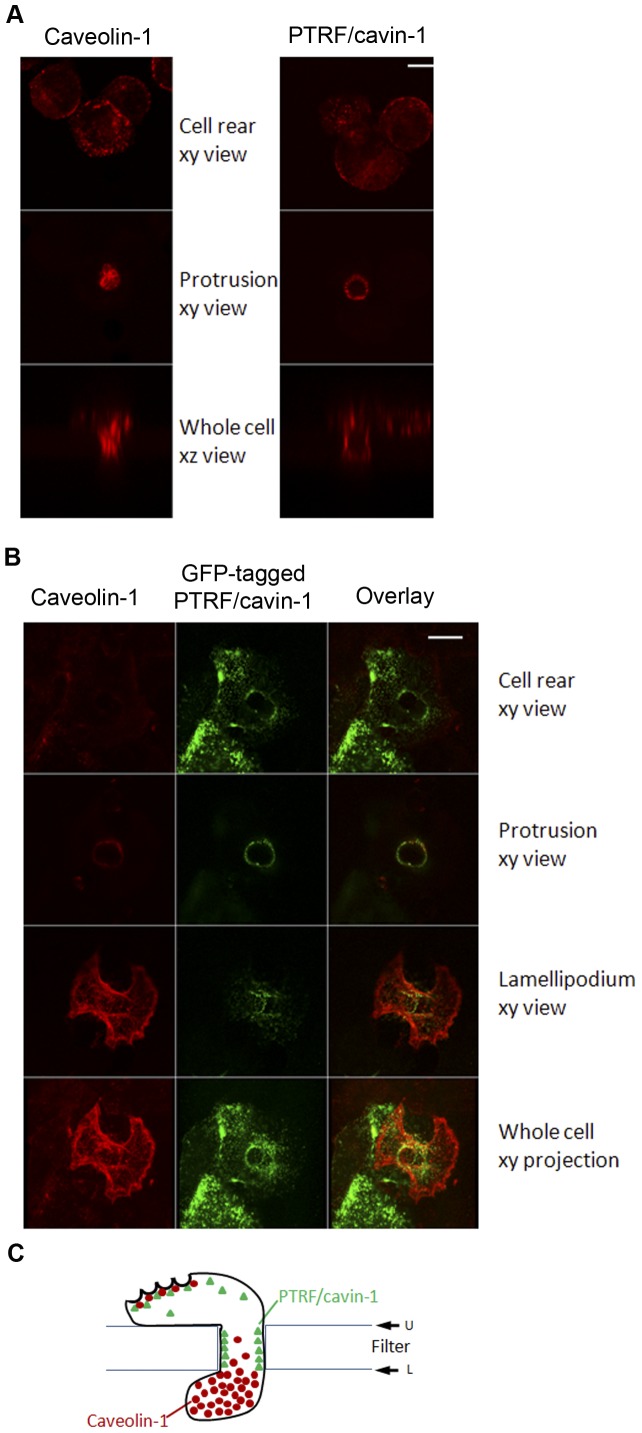
Figure 3. Differential polarization of caveolin-1 and PTRF/cavin-1 during 3D migration.
(A) Transmigrating NIH 3T3 cells were fixed and immuno-labeled using anti-caveolin-1 or anti-PTRF/cavin-1 antibodies and imaged by confocal microscopy. Shown are xy planes of cell rear, protrusion through the pore or xz planes through the center of the pore. Bar represents 10 µM. (B) Transmigrating PTRF/cavin-1-GFP expressing PC3 cells were fixed and immuno-labeled using anti-caveolin-1 antibody followed by biotinylated anti-rabbit antibody and texas red avidin. Cells were then imaged by confocal microscopy. Bar represents 10 µM. (C) Schematic representation of the differential caveolin-1 and PTRF/cavin-1 polarization during transmigration.

