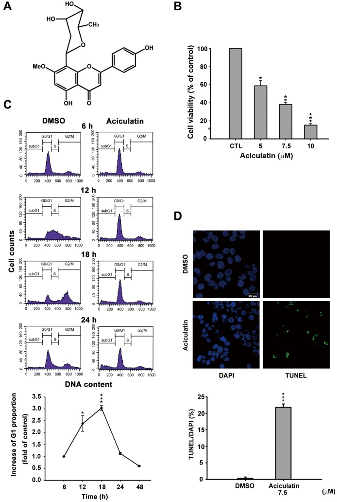
Figure 1. Effect of aciculatin on cell viability and cell cycle in human cancer cells.
A, Structure of aciculatin: 8-(2,6-dideoxy-β-ribo-hexopyranosyl)-5-hydroxy- 2-(4-hydroxyphenyl)-7-methoxy-4H-1-benzopyran-4-one sesquihydrate. B, HCT116 cells were incubated with the indicated concentrations of aciculatin (5–10 µM) for 48 h. Cell viability was then determined by MTT assay. C, HCT116 cells were starved overnight, and then incubated with vehicle (0.1% DMSO) or aciculatin (10 µM) for the indicated period. The DNA content was subsequently analyzed by PI staining using a FACScan flow cytometric assay. The curve chart shows that aciculatin increased the G1 population of cells in a time-dependent manner. Mean ± SE values from 3 independent experiments. *P<0.05 and ***P<0.001, compared with non-treated cells. D, HCT116 cells were treated with vehicle (0.1% DMSO) or aciculatin (7.5 µM) and then double stained with TUNEL and DAPI. Increased green fluorescence indicated that the cells underwent apoptosis after aciculatin treatment (TUNEL, right panel). The nuclei were stained with DAPI (left panel). The bar chart shows the proportions of TUNEL positive cells in each treatment normalized to DAPI. ***P<0.001.

