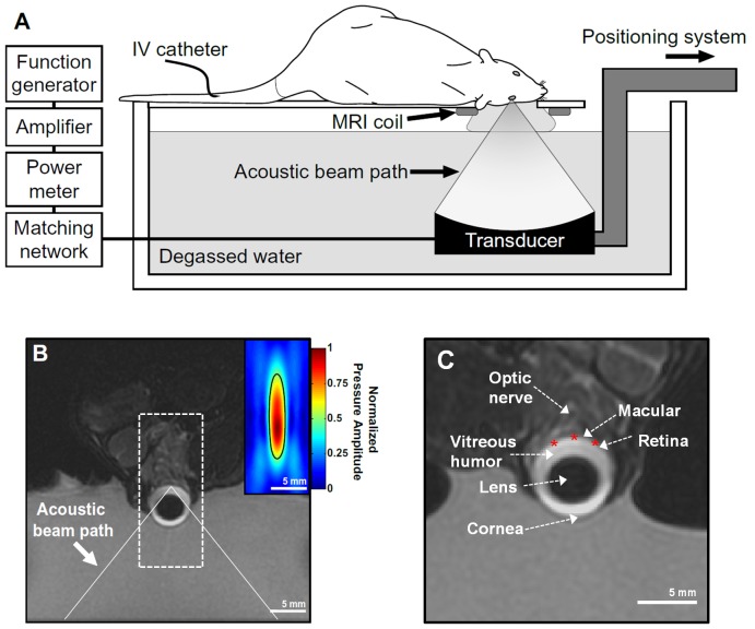
Figure 1. BRB disruption in the rat eye using an MRI-guided FUS system.
(A) Schematic diagram of the experimental set up used to disrupt the BRB. (B) Coronal T2-weighted MR image of a rat eye within the sonication system. The eye was partially submerged in water to allow for acoustic coupling. The ultrasound beam path is superimposed. (Inset) Normalized focal pressure distribution of the focal region of the ultrasound beam (boxed region) displayed at the same scale as the MR image. (C) Relevant anatomical features of the eye visible in the MR image. The location of three of the five locations targeted for sonication are shown as star symbols.

