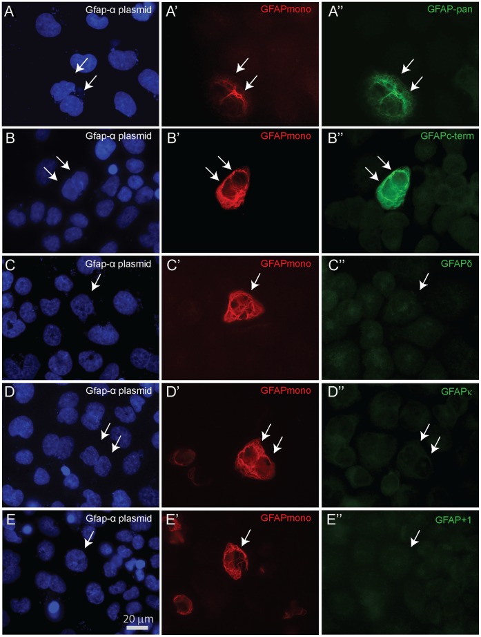
Figure 4. GFAPα transfected cells stained with various GFAP antibodies.
All panels show SW13/cl.2 cells transfected with full length msGfap-α, stained with GFAP monoclonal antibody to detect successfully transfected cells and double stained with the different polyclonals. (A–A’) GFAPpan and (B–B’) GFAPc-term antisera are able to detect GFAPα composed IF networks, whereas (C–C’) msGFAPδ, (D–D’) msGFAPκ, and (E–E’) msGFAP+1 display no reactivity against the canonical GFAPα. Panels A–E show DAPI staining, a fluorescent stain that binds to DNA.

