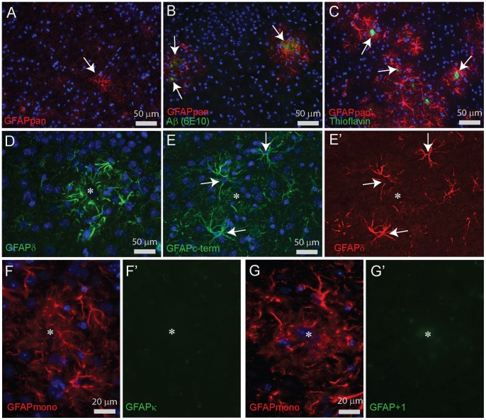
Figure 11. Immunocytochemical stainings for GFAP in cortex of APPswePS1dE9 mice.
(A) GFAPpan staining in a WT mouse cortex showing a faintly stained network of fine filaments and a single astrocyte with an intense staining (arrow). (B) GFAPpan staining in the cortex of a 6 month APPswePS1dE9 mouse with amyloid deposits (arrows), detected by Aβ staining (6E10), illustrating the close spatial association of amyloid and increased GFAP staining. (C) GFAPpan staining in the cortex of a 9 month APPswePS1dE9 mouse showing that the reactive gliosis remains associated with plaques visualized by Thioflavin-S staining (arrows). (D) GFAPδ staining is strongly enhanced in reactive astrocytes around a plaque identified by a diffuse staining in the DAPI channel (asterisk). (E,E’) Double staining of the goat polyclonal GFAPc-term and rabbit polyclonal GFAPδ demonstrating the perfect overlap of GFAPα and GFAPδ localization in reactive astrocytes (arrows) around a plaque (asterisk) in a 9 month old APPswePS1dE9 mouse. (F,F’) Double staining of GFAPmono and GFAPκ showed no GFAPκ specific staining around a plaque in a 9 month old APPswePS1dE9 mouse. (G,G’) Double staining of GFAPmono and GFAP+1 showed no GFAP+1 specific staining around a plaque in a 9 month old APPswePS1dE9 mouse.

