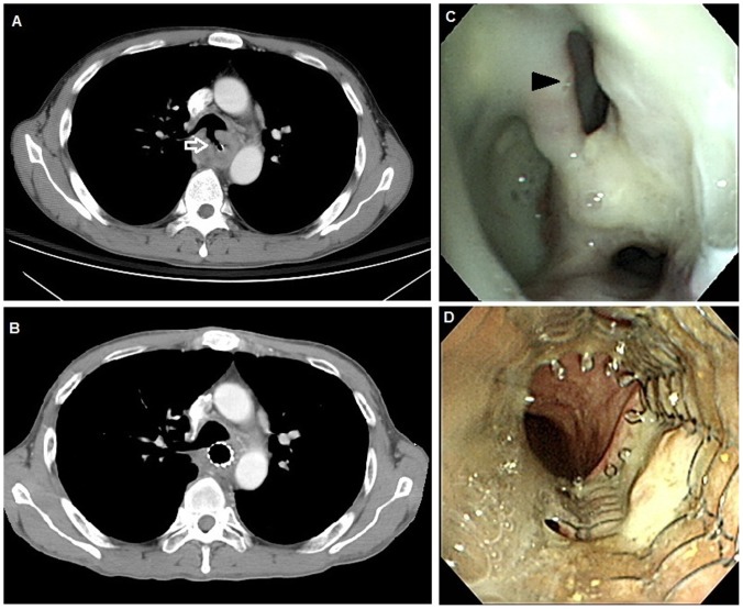
Figure 1. Representative images before and after esophageal metallic stent placement.
A. Computed tomography of the chest obtained before stent placement showed a tracheoesophageal fistula (arrow). B. Computed tomography of the chest obtained after stent placement showed a metallic stent in the esophagus covering the tracheoesophageal fistula. C. Before stent placement, endoscopic picture showed a protruding mass with a hole in the esophagus, suggesting esophageal cancer with a tracheoesophageal fistula (arrowhead). D. Endoscopic picture of an esophageal metallic stent in place one month after insertion.

