
FIGURE 5:
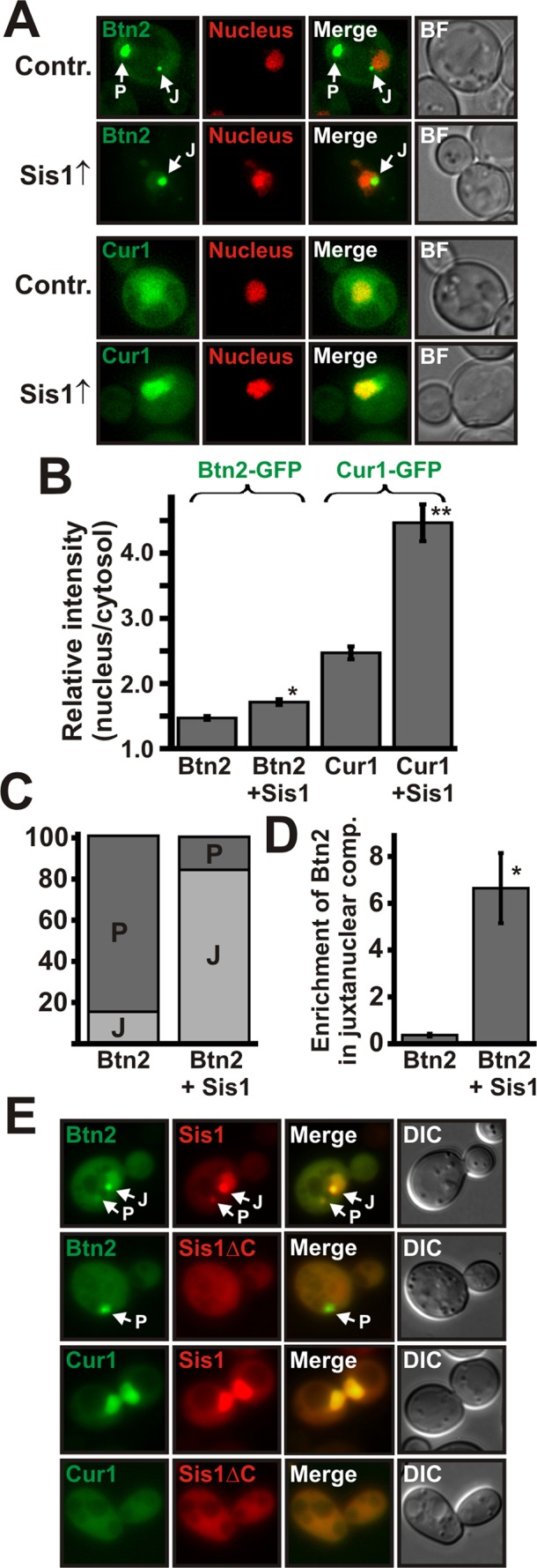
Complex formation between Sis1 and Btn2 or Sis1 and Cur1 is required for targeting to the nucleus. (A) Low-copy expression plasmids for GFP-tagged Btn2 and Cur1 were introduced into a BY4741 strain that contained a control plasmid or a low-copy expression plasmid for Sis1. Fluorescence microscopy was performed at 25°C. (B) Quantification of the relative nuclear:cytosolic GFP pixel intensity of the strains shown in A. *p = 3.3 × 10⁻⁵; **p = 2.6 × 10⁻¹⁰. (C) Quantification of the fraction of cells containing Btn2-positive juxtanuclear (J) and/or peripheral (P) foci. On the basis of the distribution of Bnt2-GFP, we arbitrarily divided cells into the two categories J and P (see Materials and Methods for details). (D) Juxtanuclear and peripheral signals were quantified (total integrated pixel intensity) in 30 cells that simultaneously contained one juxtanuclear and one peripheral compartment; p = 0.000094. (E) Chromosomal SIS1 was deleted in BY4741 yeast, and the deletion was covered with expression plasmids for mCherry-tagged SIS1 or SISΔC. Expression plasmids for GFP-tagged BTN2 and CUR1 were introduced, and the cells were observed by fluorescence microscopy.