
FIGURE 5:
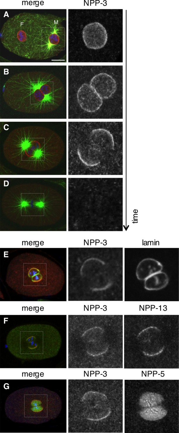
NPP-3 localization at mitotic onset. (A–D) Wild-type, one-cell-stage embryos in prophase (A and B), prometaphase (C), or metaphase (D) stained for NPP-3 (shown alone in the insets on the right and in red in the merged images), α-tubulin (green), and DNA (blue). (E–G) Wild-type, one-cell-stage embryos stained for NPP-3 (shown alone in the insets in the middle and in red in the merged images), lamin, NPP-13, or NPP-5, as indicated (shown alone in the insets on the right and in green in the merged images). DNA is shown in blue.