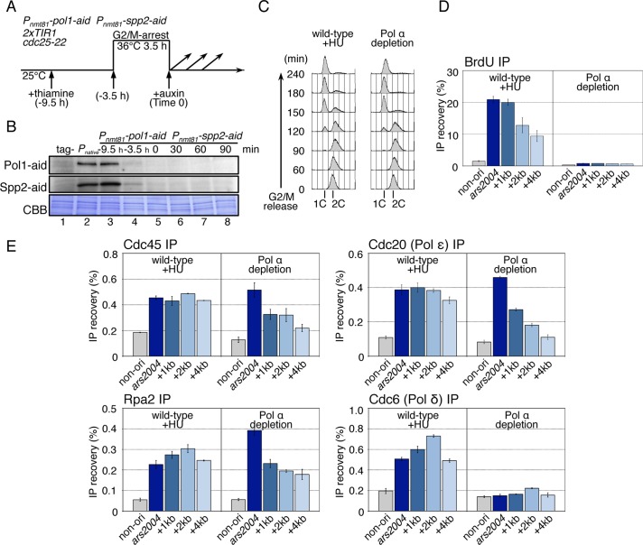
FIGURE 3:
CMG helicase and Pol ε can migrate from the origin in the absence of Pol α and DNA synthesis. (A) The experimental scheme for Pol α depletion is shown. HM3021 2xTIR1 cdc25-22 (wild-type) or the HM4011 Pnmt81-pol1-aid Pnmt81-spp2-aid 2xTIR cdc25-22 (Pol α depletion) strain grown at 25°C were cultured in the presence of thiamine (10 μg/ml) for 6 h and arrested at G2/M-phase by incubation at 36°C for 3.5 h, and then released at 25°C (Time 0). Auxin (0.5 mM) was added just before release. (B) The Pol1-aid and Spp2-aid proteins in WCE were analyzed by immunoblotting using anti-IAA17 antibody. Tag- (lane 1) and Pnative (lane 2) indicate the samples of the untagged strain (HM3021) and the strain for Pol1-aid and Spp2-aid, expressed from the native promoter (HM4031, pol1-aid spp2-aid 2xTIR1 cdc25-22) grown at 25°C, respectively. The samples of Pol α depletion (HM4011) were prepared at –9.5 h (lane 3), –3.5 h (lane 4), 0 (lane 5), 30 (lane 6), 60 (lane 7), and 90 min (lane 8) after release from G2/M. CBB represents total proteins bound on the membrane stained with Coomassie brilliant blue. (C) Wild-type (HM3696 Padh1-TK Padh1-hENT 2xTIR1 cdc25-22) and Pol α depletion (HM4111 Pnmt81-pol1-aid Pnmt81-spp2-aid Padh1-TK Padh1-hENT 2xTIR1 cdc25-22) cells were synchronously released from the G2/M boundary in the presence of BrdU (200 mM), with the addition of HU (10 mM) in the case of wild type. Aliquots taken at the indicated time points were analyzed by flow cytometry. Positions of 1C and 2C DNA contents are shown. (D) Samples taken at 90 min after G2/M release in (C) were analyzed by BrdU-IP assay as described in the legend to Figure 2B. The reproducible results obtained in biologically independent experiments are shown in Supplemental Figure S3A. (E) Wild-type and Pol α-depletion derivatives carrying flag-cdc45, cdc20-flag, or cdc6-flag were analyzed by ChIP assay at 80 min after G2/M release. In case of the wild type, HU was added at 10 mM upon G2/M release. ChIP DNAs with anti-FLAG or anti-Rpa2 were analyzed as described in the legend to Figure 2C. The reproducible results obtained in biologically independent experiments are shown in Figure 4C and Supplemental Figure S3B.

