
FIGURE 7:
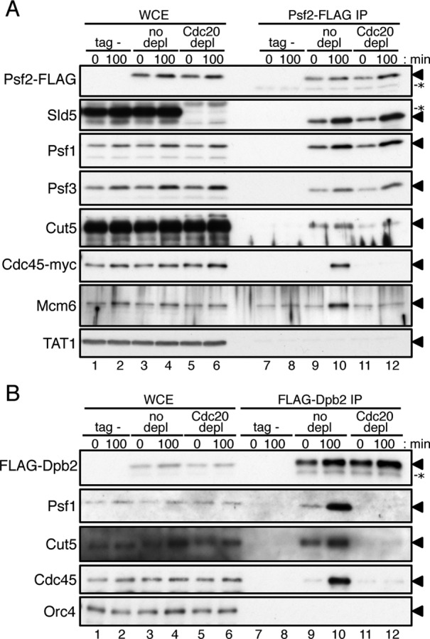
Pol ε is required for GINS to form complexes with Cut5, Cdc45, and Mcm6. Cdc20 protein was depleted as described in Figure 6. (A) Aliquots of HM3454 Pnmt81-cdc20-aid cdc45-myc 2xTIR1 cdc25-22 (tag-) and HM3450 Pnmt81-cdc20-aid psf2-flag cdc45-myc 2xTIR cdc25-22 without or with depletion (no depletion or Cdc20 depletion) were collected at 0 min and 100 min without addition of HU after G2/M release (see Figure 6) and treated with 1% formaldehyde. Proteins in WCE (lanes 1–6) and immunoprecipitates obtained with anti-FLAG (Psf2-FLAG IP, lanes 7–12) were analyzed by immunoblotting with anti-FLAG, anti-Sld5, anti-Psf1, anti-Psf3, anti-Cut5, anti-myc (Cdc45-myc), anti-Mcm6, or anti-tubulin antibodies. Triangles (◂) on the right of the panels indicate positions of the relevant proteins. Nonspecific protein bands are indicated by asterisks (*). It is noteworthy that thiamine-repressible proteins in WCE (lanes 1–4 in Sld5 panel) cross-reacted with the anti-Sld5 antibody (unpublished observation). The samples used for WCE (lanes 1–6) corresponded to 20% (Psf2-FLAG, Sld5, Psf1, Psf3, and tubulin), 1% (Cut5, Cdc45-myc), and 0.1% (Mcm6) of the proteins used for Psf2-FLAG IP. (B) Aliquots of HM4661 Pnmt81-cdc20-aid drc1-HA 2xTIR1 cdc25-22 (tag-) and HM4664 Pnmt81-cdc20-aid flag-dpb2 drc1-HA 2xTIR cdc25-22 without or with depletion (no depletion or Cdc20 depletion) at 0 min and 100 min after G2/M release were analyzed as in (A). Proteins in WCE (lanes 1–6) and immunoprecipitates obtained with anti-FLAG (FLAG-Dpb2 IP, lanes 7–12) were analyzed by immunoblotting with anti-FLAG, anti-Psf1, anti-Cut5, anti-Cdc45, or anti-Orc4 antibodies. Positions of the relevant proteins and a nonspecific protein are indicated by triangles (◂) and an asterisk (*), respectively. The samples used for WCE (lanes 1–6) corresponded to 20% (FLAG-Dpb2, Orc4) and 1% (Psf1, Cut5, and Cdc45) of the proteins used for FLAG-Dpb2 IP.