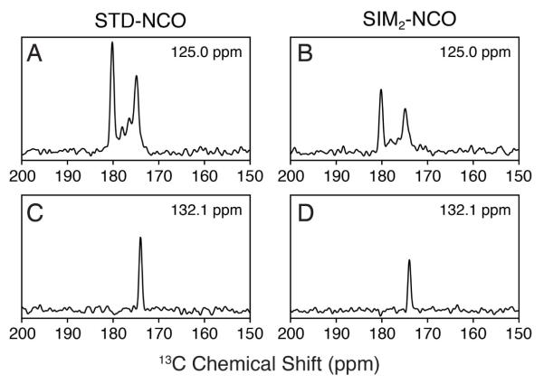
Figure 5.
1D cross sections of the 2D spectra shown in Figure 4 for the [1,3-13C] glycerol labeled ubiquitin sample. The 15N frequencies indicated in each panel are directly comparable for the STD-NCO (A, C) and SIM2-NCO spectra (B, D). The noise level is the same for the four 1D cross-sections.

