Figure 1.
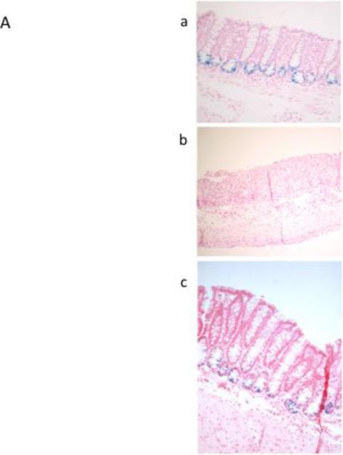
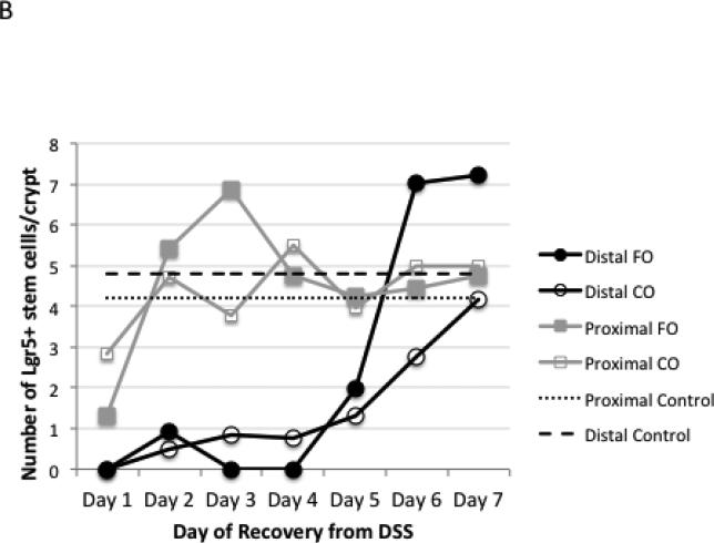
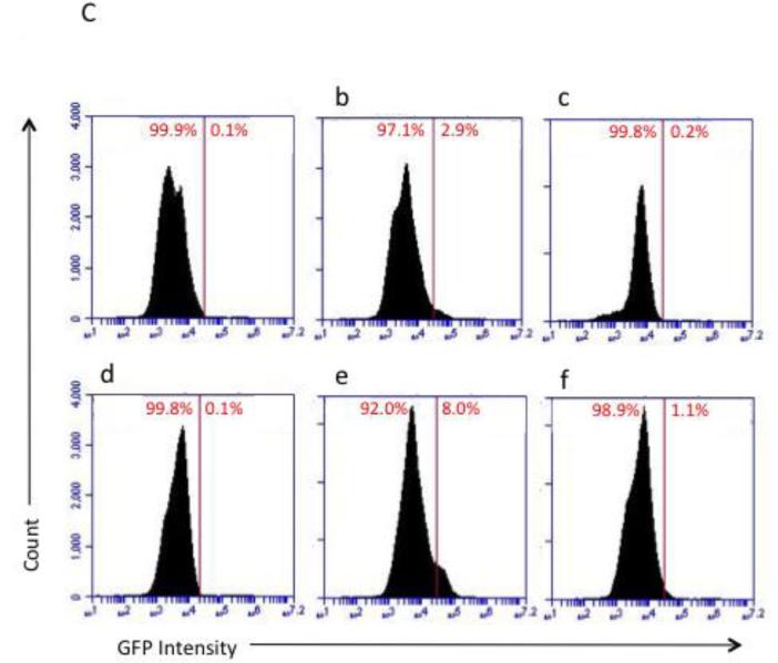
Detection of Lgr5+ stem cells during recovery from DSS treatment. Animals were treated with DSS for 5 d to induce colitis and terminated at daily intervals during the “recovery” phase. A, representative photomicrographs depicting β-galactosidase staining of Lgr5+ stem cells in distal colon in (a) untreated control, (b) DSS treated, 2 d recovery, (c) DSS treated, 7 d recovery, x100-200. B, mean number of Lgr5+ stem cells per crypt during DSS recovery (at least 20 crypts each from n=3 mice per diet group and time point. However, during days 1-4 of recovery, many animals had no scoreable crypts in the distal colon.) There were no significant differences (P>0.05) between CO and FO fed mice. C, representative flow cytometric analysis of Lgr5 GFP+ colonic epithelial cells. a-c: distal colon; d-f: proximal colon. a and d are from wild type mice (no GFP) and were used for gating. b and e represent control animals displaying the expected levels of GFP+ cells, c and f are from DSS treated mice, d 1 recovery. FO, fish oil fed; CO, corn oil fed mice.


