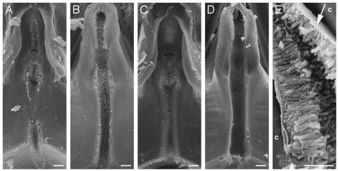
Fig. 4.
Formation of polar biofilm in insect foreguts. Scanning electron micrographs of the precibarium epipharynx (A and C) and hypopharynx (B and D) of blue-green sharpshooter leafhoppers fed on grapevines infected with the wild-type strain Temecula (A, B, and E)orthe rpfF mutant KLN61 (C and D). The stylets, which are inserted into the plant, are located above, and the cibarium (pumping chamber) is located below, the frame of the images. Xylem sap enters the precibarium from the top and runs through the canal, which is coated with a biofilm by wild-type cells (A and B) but not rpfF mutant cells (C and D). Small bits of debris are present; however, these objects were each examined at high magnification and do not represent bacterial cells, which are characteristically rod-shaped. (E) High magnification of polar biofilm that has slightly detached from cuticle (c) during fixation, revealing a mat-like structure at the attachment site (arrow). Rod-shaped structures are bacterial cells. [Bar = 10 μm (A-D) and 5 μm (E).]

